Subtotal: $31.00
51425/51415- Stoelting Tissue Slicer – Stoelting
$1,170.00 – $1,759.00
Description
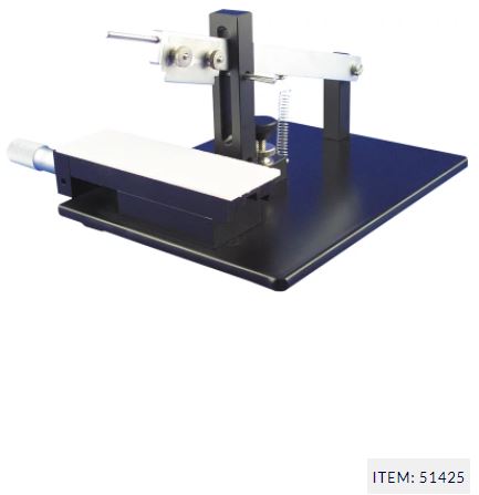
Originally designed to prepare thin slices of living brain, the tissue slicer is also excellent for slicing other tissues, blocking fixed tissue for histology, and slicing electrophoretic gels. Sections as thin as 150 microns can be reliably prepared.
Overview
Fast production of living tissue slices
Originally designed to prepare thin slices of living brain, the tissue slicer is also excellent for slicing other tissues, blocking fixed tissue for histology, and slicing electrophoretic gels. Sections as thin as 150 microns can be reliably prepared.
The tissue cutting is done with a standard double-edged razor blade.
Two Models
- The 51425 Tissue Slicer has a conventional vernier micrometer, resolution 10 microns.
- The 51415 Digital Tissue Slicer has a digital display electronic micrometer, with 1 micron resolution.
Reversible Handedness
The tissue slicer can be reversed as to left or right handedness.
Digital Tissue Slicer Easy to Use
- Switch from mm to inches
- Set zero at any point
- Set low and high tolerance limits
- Features over-travel warning signal and output to a printer, processor, data logger or computer
In use, the tissue is placed on the moistened filter paper and moved beneath the cutting edge. The cutting arm is lifted by its handle. The pin is removed and the arm is released, falling by its own weight (and assisted by the spring) thereby slicing through the tissue at high velocity. The cut tissue slice is removed from the razor blade with a small artist’s brush moistened in incubation media. The cutting arm is lifted, the dovetail slide moved for the next slice, and the process repeated.
| Specifications | |
| Distance traveled per division mark | 10 microns |
| Distance travelled per full revolution | 500 microns |
| Maximum micrometer travel distance | 25 mm |
| Dimensions (W x L x H) | 6″ x 7.5″ x 4.5″ (w/o micrometer) 8.5″ x 7.5″ x 4.5″ (with micrometer) |
| Weight | 9 lbs. (4 kg) |
| Product | 51425 (Tissue Slicer), 51415 (Tissue Slicer with Digital) |
|---|

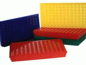 0091 Polypropylene Reversible 1.5ml And 2.0ml Microcentrifuge Tube Rack, 96 Places (Pack Of 5) Bio Plas